


Contributing € 5
Acknowledgment
Personal acknowledgment in the social networks where our group is active.
> 03 Apoiadores
We use own and third party cookies to improve your user experience and our services, analyzing users' browsing in our website. If you continue browsing, we will consider that you consent to its use. You can get further information in our Cookies Policy
You have to sign in to interact with the Goteo community
Prevención de enfermedades con Materiales Antimicrobianos
 Leganés (Madrid)


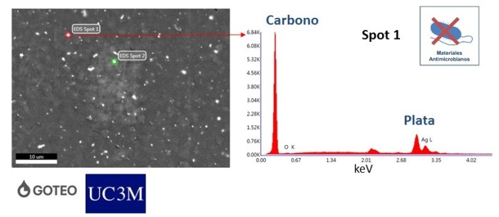
En este estudio observamos la superficie de nuestros materiales con un microscopio electrónico de barrido (también conocido por sus siglas en inglés, Scanning Electron Microscopy, SEM). Este microscopio nos permite ver con mucha resolución detalles muy pequeños en las muestras.
En este caso lo utilizamos para ver la dispersión de las nanopartículas de plata en el polietileno. La imagen muestra unos pequeños puntos brillantes que se corresponden con las partículas de plata. Para confirmarlo hicimos microanálisis de rayos-X en los puntos que están marcados en la figura. Observamos que los componentes principales de la muestra son el carbono (del polietileno) y la plata (de las nanopartículas). Concluimos que la dispersión de las nanopartículas en el material es, en general, bastante homogénea.
Inicia sesión para dejar un comentario